The Hypothalamic-Pituitary-Gonadal Axis: Molecular Coordination of Reproductive Endocrine Functions
The hypothalamic-pituitary-gonadal (HPG) axis is a central neuroendocrine system responsible for coordinating reproductive functions through interactions between the brain and the gonads. It integrates hormonal signals, neuronal activity, and peripheral physiological information to regulate sexual development, gametogenesis, steroid hormone production, and reproductive function.
This regulatory network involves three principal anatomical components: the hypothalamus, the anterior pituitary gland, and the gonads. Their coordinated activity depends on a complex sequence of hormonal interactions and feedback mechanisms that maintain reproductive endocrine regulation.
Beyond its role in reproduction, the HPG axis also interacts with metabolic, immune, and circadian regulatory systems, illustrating the interconnected nature of endocrine physiology.
1. The Hypothalamus: Initiating Reproductive Endocrine Signaling
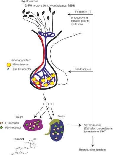
The hypothalamus acts as the principal neuroendocrine regulator of the HPG axis. Specialized neurons located primarily within the preoptic area and other hypothalamic regions synthesize and release gonadotropin-releasing hormone (GnRH).
GnRH is secreted in a pulsatile manner into the hypophyseal portal circulation, allowing it to reach the anterior pituitary gland.
The frequency and amplitude of GnRH pulses influence the synthesis and secretion of two essential gonadotropins: follicle-stimulating hormone (FSH) and luteinizing hormone (LH).
This pulsatile organization is essential because continuous exposure to GnRH can lead to reduced pituitary responsiveness through receptor desensitization and downregulation.
Hypothalamic GnRH secretion is also influenced by several regulatory signals, including kisspeptin, neurokinin B, dynorphin, metabolic hormones, and environmental factors.
Kisspeptin-producing neurons are particularly important because they provide a major stimulatory input to GnRH neurons through the KISS1 receptor.

2. The Anterior Pituitary: Gonadotropin Synthesis and Release
Following GnRH stimulation, gonadotroph cells in the anterior pituitary synthesize and release FSH and LH.
Both hormones belong to the glycoprotein hormone family and share a common alpha subunit, while their distinct beta subunits confer biological specificity.
FSH primarily regulates processes associated with gamete development and supporting cell activity.
In females, FSH stimulates the growth and maturation of ovarian follicles by acting on granulosa cells. It also promotes aromatase expression, an enzyme involved in converting androgens into estrogens.
In males, FSH acts on Sertoli cells within the seminiferous tubules, supporting spermatogenesis and the development of germ cells.
LH performs complementary functions by stimulating steroid hormone production.
In females, LH acts on ovarian theca cells to promote androgen synthesis, which provides substrates for estrogen production in granulosa cells. It also participates in ovulation and corpus luteum formation.
In males, LH stimulates Leydig cells to produce testosterone, an essential hormone for male reproductive physiology.

3. Gonadal Function: Hormone Production and Gametogenesis
The gonads represent the peripheral component of the HPG axis and respond to pituitary gonadotropins through specialized hormone receptors.
Their functions include the production of reproductive cells and the synthesis of steroid hormones.
Testicular endocrine activity
Testicular function depends on interactions between Leydig cells, Sertoli cells, and developing germ cells.
LH stimulates Leydig cells to synthesize testosterone through steroidogenic pathways involving cholesterol transport and mitochondrial enzymatic activity.
FSH acts on Sertoli cells, supporting germ cell development, the formation of the blood-testis barrier, and the production of regulatory proteins such as inhibin B.
Testosterone and FSH therefore contribute through complementary mechanisms to the maintenance of spermatogenesis.
Ovarian endocrine activity
Ovarian function involves coordinated interactions between granulosa cells, theca cells, and developing oocytes.
During follicular development, FSH promotes granulosa cell proliferation and estrogen synthesis, while LH stimulates androgen production in theca cells.
The conversion of androgens into estradiol through aromatase activity is a central component of ovarian steroidogenesis.
Following ovulation, the corpus luteum produces progesterone and additional estrogen, contributing to the hormonal regulation of the reproductive cycle.
4. Steroidogenesis: The Molecular Basis of Reproductive Hormone Production
Steroidogenesis is the biochemical process through which steroid hormones are synthesized from cholesterol.
It occurs primarily in specialized endocrine cells of the adrenal glands and gonads.
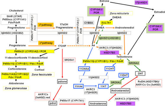
Within gonadal tissues, cholesterol is transported into mitochondria through the steroidogenic acute regulatory protein (StAR). This step facilitates its conversion into pregnenolone by the cytochrome P450 side-chain cleavage enzyme (CYP11A1).
Pregnenolone subsequently undergoes several enzymatic transformations involving proteins such as CYP17A1, 3β-hydroxysteroid dehydrogenase, and 17β-hydroxysteroid dehydrogenase.
These reactions contribute to the production of testosterone, estradiol, and progesterone.
The relative expression and activity of steroidogenic enzymes vary according to cell type, hormonal stimulation, and physiological context.
Steroid hormone production is therefore closely connected to the regulatory activity of the HPG axis.
5. Feedback Mechanisms: Maintaining Endocrine Equilibrium
The HPG axis operates through sophisticated feedback mechanisms that regulate hormonal secretion and maintain physiological stability.
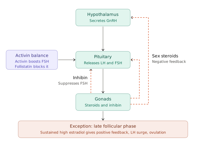
Negative feedback occurs when circulating sex steroids reduce hypothalamic GnRH activity and pituitary gonadotropin secretion.
In females, estradiol and progesterone generally exert negative feedback during much of the reproductive cycle. In males, testosterone and its metabolites contribute to the regulation of GnRH and gonadotropin release.
Inhibin, produced by ovarian granulosa cells and testicular Sertoli cells, selectively suppresses FSH secretion at the pituitary level.
Activin has an opposing stimulatory effect on FSH synthesis, while follistatin binds activin and reduces its biological activity.
An important exception to the predominantly negative feedback pattern occurs during the late follicular phase of the female reproductive cycle. Sustained high estradiol concentrations trigger positive feedback, producing the preovulatory LH surge that initiates ovulation.
These mechanisms demonstrate how hormonal feedback adapts endocrine activity to changing physiological requirements.
6. Molecular Signaling Pathways Within the HPG Axis
The biological activity of reproductive hormones depends on intracellular signaling pathways that translate receptor activation into cellular responses.
GnRH receptors are G protein-coupled receptors that primarily activate phospholipase C signaling. This leads to the production of inositol trisphosphate (IP3) and diacylglycerol (DAG), intracellular messengers that regulate calcium mobilization and protein kinase C activity.
FSH and LH receptors also belong to the G protein-coupled receptor family. Their activation commonly stimulates adenylate cyclase, increasing intracellular cyclic AMP (cAMP) and activating protein kinase A (PKA).
These signaling mechanisms regulate transcription factors, steroidogenic enzymes, cell proliferation, and hormone synthesis.
Additional pathways, including MAPK/ERK and PI3K/AKT, contribute to gonadal cell responses and interactions between endocrine signals and cellular metabolism.
The integration of these pathways enables reproductive tissues to respond to hormonal information with appropriate molecular and physiological changes.
7. Metabolic and Environmental Regulation of the HPG Axis
Reproductive endocrine activity is influenced by physiological conditions extending beyond the reproductive organs.
Energy availability, nutritional status, stress responses, sleep patterns, and circadian rhythms can modify HPG axis activity.
Leptin, insulin, and other metabolic signals provide information about the body's energy status and influence hypothalamic regulatory networks, particularly kisspeptin-related mechanisms.
Stress-related activation of the hypothalamic-pituitary-adrenal axis can also interact with reproductive regulation through glucocorticoids and other neuroendocrine mediators.
Circadian organization contributes to the timing of hormonal secretion and reproductive physiological processes.
These interactions demonstrate that reproductive regulation is integrated with broader systems responsible for maintaining physiological adaptation.
8. Research Approaches for Investigating HPG Axis Regulation
The investigation of the HPG axis requires complementary analytical approaches capable of evaluating hormonal concentrations, molecular expression, and cellular responses.
Common research techniques include:
Immunoassays: ELISA and related methods are used to quantify circulating GnRH-related markers, FSH, LH, estradiol, progesterone, testosterone, and inhibin.
Molecular analysis: RT-qPCR allows the investigation of genes involved in hormone synthesis, receptor expression, and endocrine regulation.
Protein analysis: Western blotting and immunohistochemistry help characterize hormone receptors, steroidogenic enzymes, and intracellular signaling components.
Cellular models: Specialized gonadal cell models can be used to examine responses to gonadotropins and other endocrine signals.
Advanced imaging: Microscopy techniques support the investigation of tissue organization, cellular interactions, and protein localization.
9. Emerging Perspectives in Reproductive Neuroendocrinology
Contemporary research increasingly explores the HPG axis as part of a broader network connecting neural activity, endocrine regulation, metabolism, and immune function.
Single-cell transcriptomics, advanced molecular profiling, and computational modeling are helping researchers investigate cellular heterogeneity within hypothalamic and gonadal tissues.
New research directions also include the molecular regulation of reproductive aging, neuroendocrine plasticity, hormone receptor sensitivity, and interactions between reproductive and metabolic pathways.
These approaches provide opportunities to investigate the complexity of reproductive regulation at increasingly precise biological levels.
An Integrated System of Neuroendocrine Coordination
The hypothalamic-pituitary-gonadal axis represents a fundamental regulatory system connecting the central nervous system with reproductive endocrine organs.
Through the coordinated release of GnRH, FSH, and LH, together with gonadal steroid production and feedback regulation, the HPG axis controls essential aspects of reproductive physiology.
Its regulation depends on interactions between hormonal signals, intracellular pathways, metabolic conditions, and environmental influences.
Understanding this integrated neuroendocrine architecture provides an important foundation for exploring reproductive biology, cellular communication, endocrine physiology, and human development.